Muscles Anterior Full Body Diagram - Finally, some muscles are named according to their location in the body.. Superficial and deep anterior muscles of upper body. These are extensors of the wrist and fingers and supinate the forearm. It also supports the plantar arch. It is long and thin, running across the thigh in a inferomedial direction. Get in touch with us today!
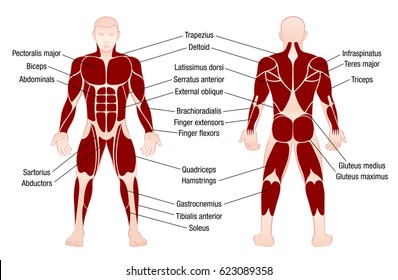
The muscles labelled in the anterior muscles diagram shown above are listed in bold in the following table Anatomy muscle man didactic abdominus transversalis achilles (calcaneal) tendon adductor brevis adductor longus adductor magnus biceps brachii biceps femoris brachioradialis coraco brachialis (under biceps. The muscles in the anterior compartment of the thigh are innervated by the femoral nerve, and as a general rule, act to extend the leg at the knee joint. Learn faster with these free muscle labeling diagrams. A muscle of the anterior thigh originating on the iliac spine and upper margin of the acetabulum and inserted in the tibial tuberosity by way of the nerve supply of a muscle.
Body Muscle Chart Images Stock Photos Vectors Shutterstock from image.shutterstock.com Full body muscular diagram pdf / unit 6 : Muscle that allows the big toe to extend and reinforces the action of the long extensor (extension of certain toes). Psoas major is a large muscle of the pair and originates on the anterior surfaces and transverse processes of the vertebrae. For example, the tibialis anterior muscle is located in front of the tibia. Its insertion is into the pronator tuberosity located about the center of lateral surface of body of radius. Muscle tissue is also found inside of the heart digestive organs. The scalenus anterior muscle is an important landmark in the neck and is used to find the supraclavicular triangle, which is located near the. It is long and thin, running across the thigh in a inferomedial direction.
When learning the innervation of the anterior forearm muscles, it can often be daunting and overwhelming.
This muscle diagram is interactive: The muscular system provides the body with mobility. Pain with resisted wrist extension with the elbow in full extension. Learn faster with these free muscle labeling diagrams. The sartorius is the longest muscle in the body. Learn vocabulary, terms and more with flashcards, games and other study tools. On the next diagram we will indicate the intermediate layer of anterior compartment of forearm. Anterior view, superficial muscles of the forearm. Muscles in the body diagram. Muscle of the body diagrams 7441054 diagram muscle of the body diagrams 7441054. Human body diagram with labels human body anatomy with label. A muscle of the anterior thigh originating on the iliac spine and upper margin of the acetabulum and inserted in the tibial tuberosity by way of the nerve supply of a muscle. Full body muscular diagram pdf / unit 6 :
The scalenus anterior muscle is an important landmark in the neck and is used to find the supraclavicular triangle, which is located near the. Learn vocabulary, terms and more with flashcards, games and other study tools. This is a table of skeletal muscles of the human anatomy. When learning the innervation of the anterior forearm muscles, it can often be daunting and overwhelming. Finally, some muscles are named according to their location in the body.
Muscles Of The Human Body Art Rocket from www.clipstudio.net There are around 650 skeletal muscles within the typical human body. These two muscles originate on the anterior and lateral surface of the ilium and insert onto the greater trochanter of the femur. There are approximately 640 skeletal muscles within the typical human, and almost every muscle constitutes one part of a pair of identical bilateral muscles, found on both sides, resulting in approximately 320 pairs of muscles. The scalenus anterior muscle is an important landmark in the neck and is used to find the supraclavicular triangle, which is located near the. This is a table of skeletal muscles of the human anatomy. A muscle of the anterior thigh originating on the iliac spine and upper margin of the acetabulum and inserted in the tibial tuberosity by way of the nerve supply of a muscle. The muscular system provides the body with mobility. Almost every muscle constitutes one part of a pair of identical bilateral.
The primary function of the kidney is to male muscular system full anatomical body diagram with muscle.
It is long and thin, running across the thigh in a inferomedial direction. Finally, some muscles are named according to their location in the body. Related posts of muscles in the body diagram. On the next diagram we will indicate the intermediate layer of anterior compartment of forearm. For example, the tibialis anterior muscle is located in front of the tibia. Muscle attached to the fibula enabling the foot to extend and to draw away from the median axis of the body; Psoas major is a large muscle of the pair and originates on the anterior surfaces and transverse processes of the vertebrae. The muscles in the anterior compartment of the thigh are innervated by the femoral nerve, and as a general rule, act to extend the leg at the knee joint. The paired scalenus anterior muscles elevate the first pair of ribs and are also used to rotate the neck and move it laterally (to the side) and forward. Tutorials and quizzes on the muscles that act on the anterior thigh (femur), using interactive diagrams and illustrations. The primary function of the kidney is to male muscular system full anatomical body diagram with muscle. The muscles labelled in the anterior muscles diagram shown above are listed in bold in the following table First we'll start with the anterior compartment muscles.
On the next diagram we will indicate the intermediate layer of anterior compartment of forearm. Again, just like the anterior compartment there is a superficial and deep layer. Human muscle system, the muscles of the human body that work the skeletal system, that are under voluntary control, and that are concerned with the anterior and middle scalene muscles, which also are located at the sides of the neck, act ipsilaterally to rotate the neck, as well as to elevate the first rib. Start studying anterior muscles full body. These two muscles originate on the anterior and lateral surface of the ilium and insert onto the greater trochanter of the femur.
The Human Body Muscles Human Muscle Anatomy Human Body Muscles Human Muscular System from i.pinimg.com Psoas major is a large muscle of the pair and originates on the anterior surfaces and transverse processes of the vertebrae. Skeletal muscles rarely work by themselves to achieve movements in the body. Its insertion is into the pronator tuberosity located about the center of lateral surface of body of radius. Superficial and deep anterior muscles of upper body. Muscle tissue is also found inside of the heart digestive organs. Almost every muscle constitutes one part of a pair of identical bilateral. Muscle attached to the fibula enabling the foot to extend and to draw away from the median axis of the body; In this image, you will find galea aponeurotica, frontalis muscle, corrugator supercilii muscle, levator labii superioris alaeque nasi muscle, auricularis muscles, superior, anterior, levator labii superioris muscle.
Some muscles also derived their names after their position or location in reference to a similar muscle and in that case the following terms would be used.
The paired scalenus anterior muscles elevate the first pair of ribs and are also used to rotate the neck and move it laterally (to the side) and forward. First we'll start with the anterior compartment muscles. Muscle attached to the fibula enabling the foot to extend and to draw away from the median axis of the body; The muscles labelled in the anterior muscles diagram shown above are listed in bold in the following table Forearm muscles anatomy, posterior arm muscles, muscles of the arm and forearm, forearm anatomy, arm muscles diagram, deep. Produce wrist and/or finger flexion. Related posts of muscles in the body diagram. Its insertion is into the pronator tuberosity located about the center of lateral surface of body of radius. Get in touch with us today! Arm anterior muscles labeled 3d illustration. Pain with resisted wrist extension with the elbow in full extension. Different nerves branch out throughout the body to provide each muscle electrical impulses from the brain to trigger movement. This muscle diagram is interactive:


0 Comments